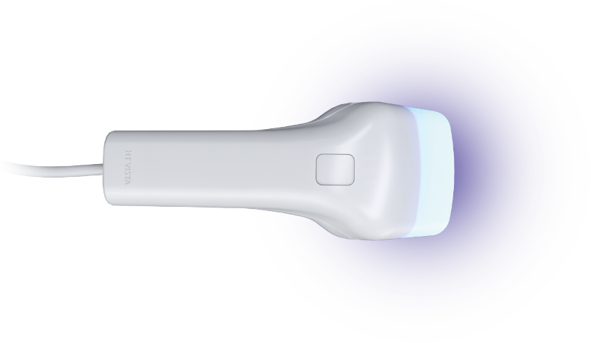
HT Vista Can Make for Your Veterinary Clinic!
A non-invasive cancer screening tool for canine dermal and sub-cutaneous masses.
How does
HT Vista
work?

-
IdentifyRegion of concern and clip a small patch of fur.
-
ScanHeat waves are sent to this tissue.
The thermal sensor measures the heat diffusion signals. -
MarkThe area of concern and healthy area.
-
Get HT Vista ReportResults delivered on-the-spot.

Non-Invasive & Pain Free
A needle free platform ensures a stress and pain free experience for the patient.
.

AVG of 98% NPV
Experience peace of mind when communicating a benign result to the pet owners.

40 Second Scan
Real-time analysis provides immediate results and facilitates discussion on next steps.

Cost Effective
A cost-effective screening tool to add to your diagnostic offerings.
Dr Michael Morris
Southwest Hospital, USA
Dr Michael Morris, a Veterinary medicine practitioner of 54 years discusses how the HT Vista has improved his clinics patient care, team development.
"I am very satisfied with the product and like the fact my team uses a cutting edge technology".
Dr Monika Wlodarchak
Tender Touch, USA
Dr Monika Wlodarchak demonstrates the importance the HT Vista plays in discovering a MCT amongst previously diagnosed Lipomas, which illustrates the benefit of introducing a screening tool into your mass investigation routine.
"We immediately took the HT Vista and scanned it, and, unfortunately, we found that it was something that needed further investigation. Underneath the original Lipoma was an invading Mast Cell Tumour. We saved the patient's life, by not ignoring that Lipoma, all because the HT Vista detected it for us."
Dr Carolyn Kutzer
Freed Hospital, USA
Dr Carolyn Kutzer agrees that the 'Wait and see' approach is not recommended, and in her experience the HT Vista proves that point. See how scanning Lipomas with the HT Vista is worth the consideration.
"This has been a huge game changer for us, showing that our guesses aren't always correct".